What Type Of Epithelium Lines The Oral Cavity
Histology membrane mucous gingiva mucosa keratinized epithelium keratinised epithelial Oral barrier mucosal tissue cell immunity immunology mucosa figure gingival specific squamous attachment Mucous epithelium pictorial
3 Oral epithelium | Pocket Dentistry
Shows a cross section of normal buccal mucosa illustrating the Tissue epithelium stratified epithelial nonkeratinized squamous function cuboidal structure location cells keratinized columnar simple non where types different found esophagus Healthy tissues and the oral cavity
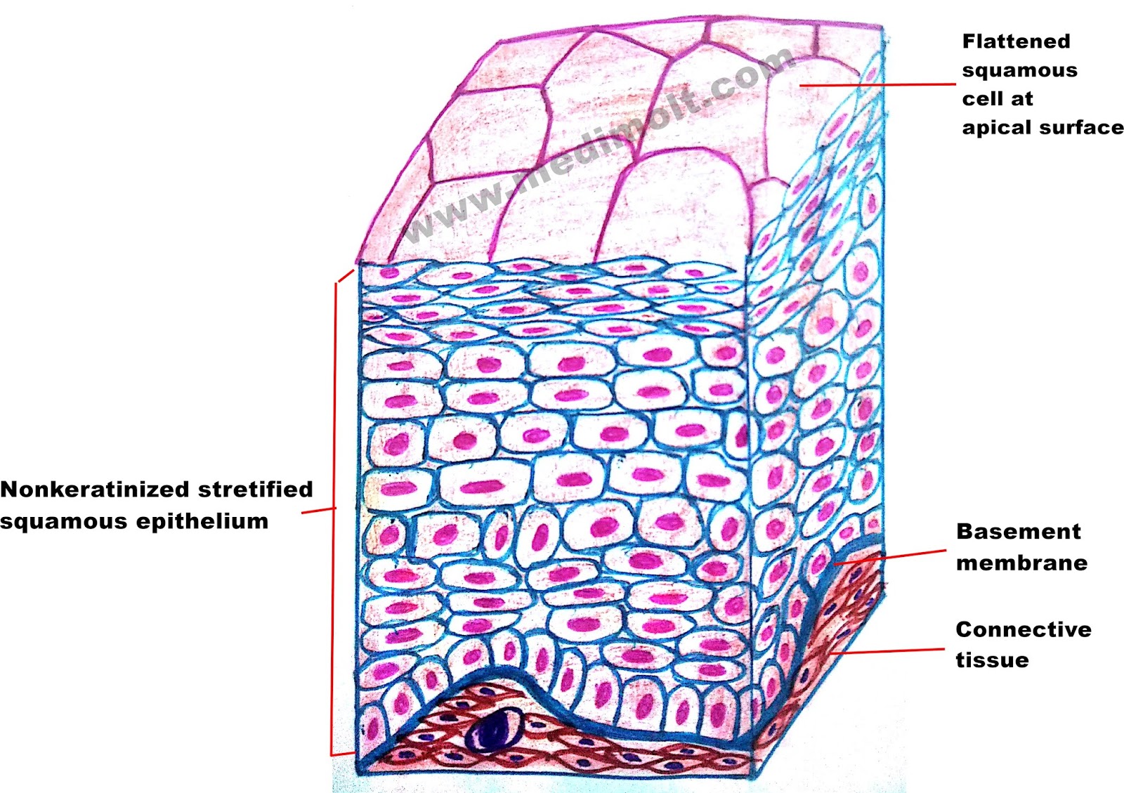
Schematic drawing illustrating different layers of the oral epithelium
Healthy tissues and the oral cavityEpithelium layers number based cells classification anatomy Oral cavityMucosa buccal cell illustrating.
What is epithelial tissue different types of structure location andEpithelium cavity keratinised Histological (left) and schematic (right) image of the buccal oralBuccal mucosa histological schematic pathology.

Oral mucosae
Tissue-specific immunity at the oral mucosal barrier: trends in immunologyEpithelium cavity Histology oral cavity glands salivary mucosa gland palate pharynx specialized stomach esophagus mucousCavity epithelium inspect.
Inspect oral cavity patterns with narrow band imaging (nbi)Oral mucosa layers epithelium lamina propria skin mouth cavity squamous lining submucosa mucosae stratified composed features figure histological tissues has Epithelium illustrating mucosa buccal servierOral tissues layers micrograph structures mucosa cavity lamina submucosa propria muscle components present light bone.
1: oral structures and tissues
Epithelium oral dentistry pocket squier finkelstein mosby 2003 copyrightOral mucous membrane pictorial representation Buccal histological mucosa pathology3 oral epithelium.
Histological (left) and schematic (right) image of the buccal oralHistology of oral mucous membrane and gingiva .


1: Oral Structures and Tissues | Pocket Dentistry

Oral Mucosae - Hot Russian Teens

Inspect Oral Cavity Patterns With Narrow Band Imaging (NBI) - Olympus

3 Oral epithelium | Pocket Dentistry

Histological (left) and schematic (right) image of the buccal oral

Schematic drawing illustrating different layers of the oral epithelium

Epithelium - Anatomy QA

Shows a cross section of normal buccal mucosa illustrating the

Healthy Tissues and the Oral Cavity - Revise Dental
